

The Eye
A collection of videos and experiments that are suitable for Biology.
How the Body Works : Exploring the Eye
As we explore the eye, we see that the eyes are organs of sight, situated in orbits, the sockets in the skull, the walls of which protect them from injury. The eyelashes, eyelids, muscles and lacrimal glands also protect these vital and delicate organs. The eye is divided into two segments by the lens and ciliary body. The front segment contains the fluid aqueous humor, and is in turn divided, by the iris, into anterior and posterior chambers, which are connected through the pupil's aperture in the iris. The back segment, called the vitreous body, contains a jellylike substance, known as the vitreous humor, and is lined by the light sensitive retina. The iris is an adjustable diaphragm with an aperture, the pupil, in its center. It acts like a valve, controlling the amount of light entering the eye. The lens is a transparent, biconvex body, enclosed in a thin, elastic, transparent capsule. It is supported by ligaments attached to the ciliary body, which can change its shape. The ciliary muscle, composed of smooth muscle under involuntary control, alters the shape of the lens. The suspensory ligaments connect the ciliary muscle to the lens, and hold the lens in place. The hyaloid canal is the remains of a channel that carried an artery during the development of the eye in the fetus. The cornea, aqueous humor, lens and vitreous humor are all transparent, thus allowing the unobstructed passage of light from the exterior, through the eyeball, to the retina. The cornea is the most important structure for refracting light, although the lens provides the fine control needed to converge the incoming rays into the retina. The most striking external feature of the eye is the iris, the pigment-filled membrane that gives the eye its color, varying from light blue to dark brown.
The focusing mechanism of the eye consists of the lens, which is completely encircled by the ciliary muscles and attached to them by the suspensory ligaments. In distant vision the ciliary muscles are relaxed and the lens is pulled flat. It does little focusing because the almost parallel light rays from the distant object need to be only slightly refracted to bring them to a point on the retina. In near vision, the ciliary muscles contract, the suspensory ligaments loosen and the lens becomes more convex. The curved surface of the lens reinforces the cornea's focusing of the more divergent rays from the near object.
Check out our most popular games!
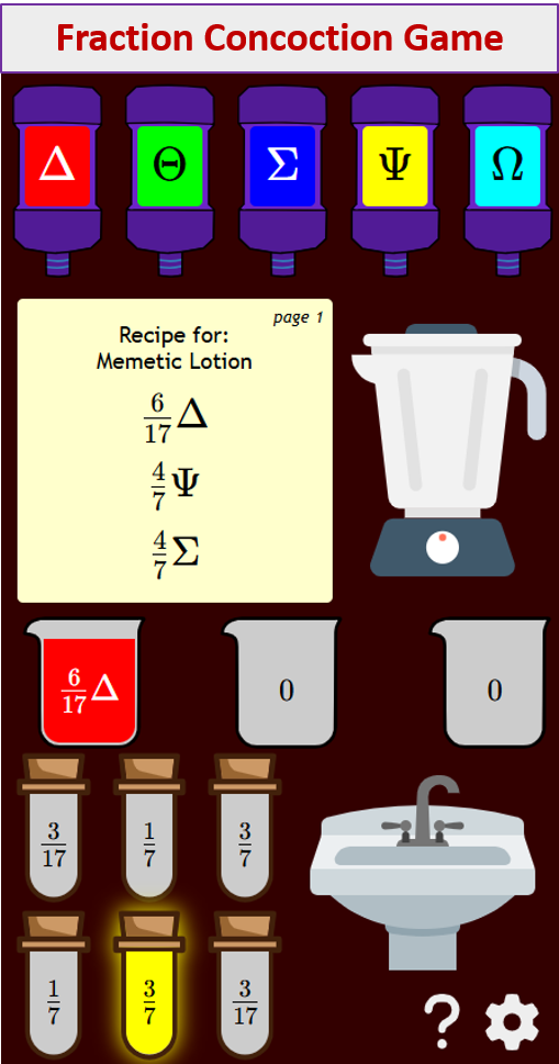
Fraction Concoction Game:
Master fractions in the lab: mix, add, and subtract beakers to create the perfect concoction!
Fact Family Game:
Complete fact families and master the link between addition & subtraction and multiplication & division.

Number Bond Garden:
Clear the board by matching number pairs that sum to ten in this garden-themed mental math puzzle.

Online Addition Subtraction Game:
Practice your addition and subtraction skills to help the penguin find its mummy.

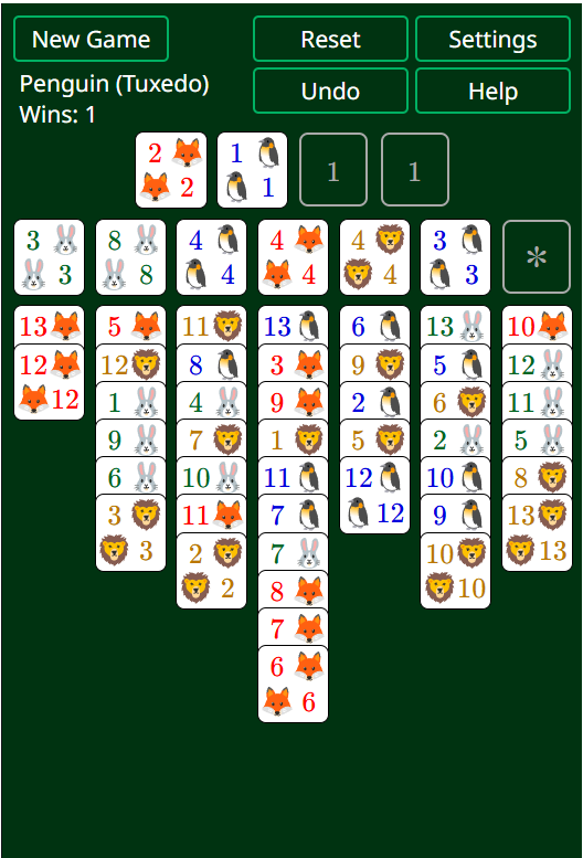
Penguin Solitaire
Penguin Solitaire is a fun game that aims to move all cards to the foundations to build four full sequences. There are two versions here: Penguin (Tuxedo) and Penguin (Original).
We welcome your feedback, comments and questions about this site or page. Please submit your feedback or enquiries via our Feedback page.


